Chronic Wound
A chronic wound develops when any acute wound fails to heal in the expected time frame for that type of wound, which might be a couple of weeks or up to six weeks in some cases.A wound that has failed to heal in 4 weeks is defined as a chronic wound.
Risk Factors; Age, immune status, malnutrition, infection, insufficient oxygenation or perfusion, smoking, diseases, medications, radiation, and chemotherapy are the main risk factors.
Types of Chronic Wounds
Some of the most common types of wounds and their specific causes are as follows:
Ulcers (the most common type of chronic wounds)
These can occur from hypertension, atherosclerosis (plugging) and thrombosis (clotting), where the reduced blood supply leads to an ischemic state.
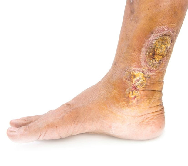
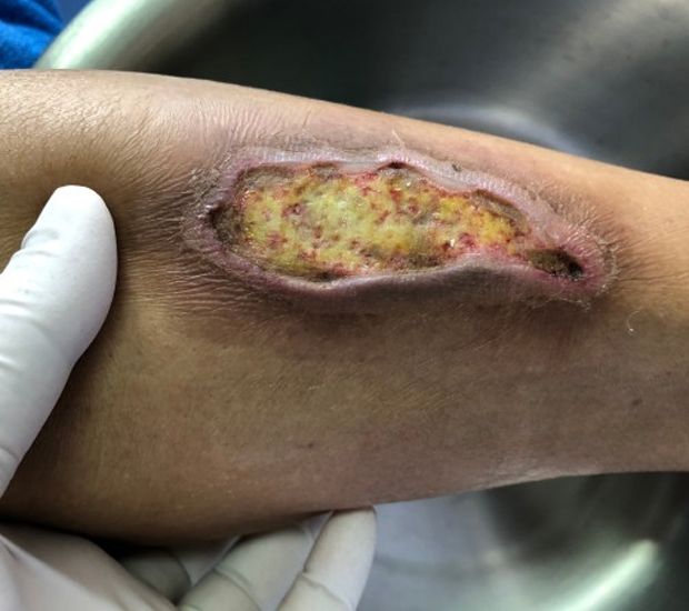
These account for more than half of ulcer cases, especially in the lower limbs (mainly the legs) as associated with deep vein thrombosis, varicose veins, and venous hypertension. Venous ulcers can lead to stasis, where the blood fails to circulate normally.
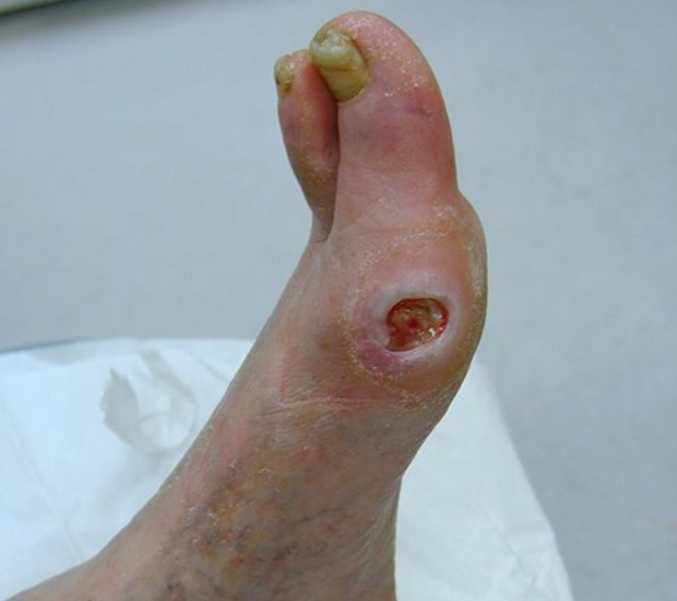
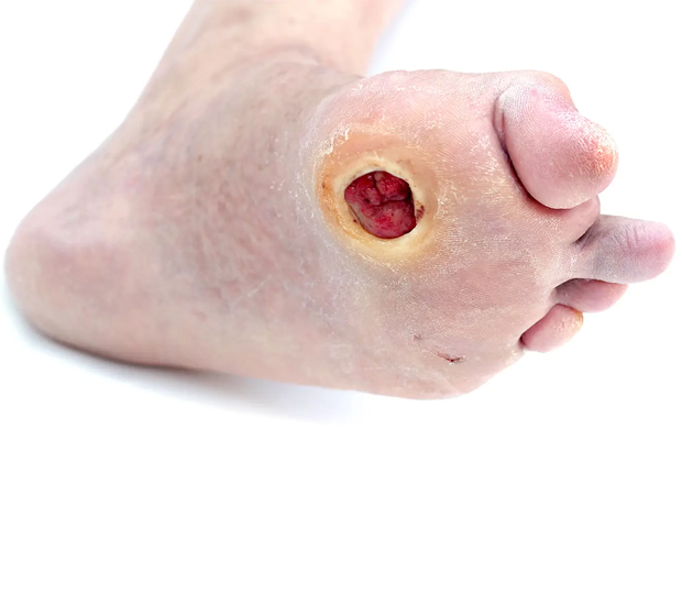
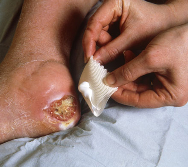
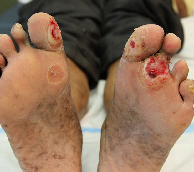
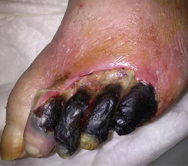
These are a common complication in uncontrolled diabetes mellitus, resulting in impaired immune function, ischemia (due to poor blood circulation) and neuropathy (nerve damage), which eventually lead to breakage of skin and ulceration.A diabetic foot ulcer is a common complication of diabetes mellitus. An ulcer will develop if there is a break in the skin which does not progress to heal, due to the underlying diabetic condition. Diabetic foot ulcers require careful management and we have a range of products which can help to manage these ulcers.
The constant pressure and friction resulting from body weight over a localized area for prolonged duration can lead to breakage of skin and ulceration (also known as bed sores); especially on the back and on the ankles and feet.
Whether it is bacterial, fungal, or viral, if the cause of the infection is not treated with the proper medication, the wound will not heal properly in the expected time.
Ischemia means that the wound area is not getting sufficient blood supply. Limiting the blood supply, and the oxygen and nutrients it carries, can delay the healing process, or even prevent it.
Regardless of whether the source of radiation was therapeutic (gamma rays or x-rays) or accidental (exposure to radioactive materials from nuclear plant accidents or radioactive devices that detonate), excessive exposure to ionizing radiating materials can weaken the immune system, cause damage to exposed tissue and delay the healing time of all wounds.
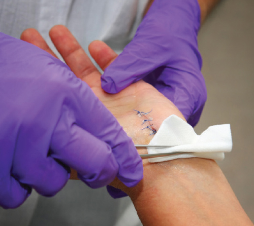
Wounds caused by incisions made during surgery can progress to chronic wounds if the blood supply to the surgery area was accidentally damaged or if wound care was inadequate. Both can delay the healing time of a wound.
Management of Chronic Leg Ulcers
The management of leg ulcers should include a detailed wound assessment, investigations, wound debridement, and treatment. Successful management of leg ulcers requires a clear diagnosis, establishment of a treatment plan, accurate monitoring, and adherence to the plan as the ulcer decreases in size. Education and training is vital for all those involved in caring for patients with chronic ulceration.
- If ulcer is infected it will be treated with antibiotics to avoid further complications.
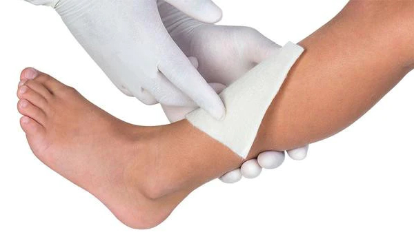
- Compression bandages are also used to help ease swelling, close the wound, and prevent infection.
- Specific ointments may be to applied to the ulcer.
- In severe cases orthotics or braces to improve gait pattern and prevent future ulcers.
Wound management products
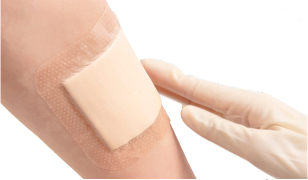
Passive dressings :
Use the ‘passive’ or the ‘plug and conceal’ concept, including gauze, lint, non-stick dressings, and tulle dressings. These products fulfil very few of the properties of an ideal dressing and have very limited, if any, use as primary dressing, but some are useful as secondary dressings.
Interactive dressings :
These dressings help to control the micro-environment by combining with the exudate to form either a hydrophilic gel or, by means of semipermeable membranes, controlling the flow of exudate from the wound into the dressing. They may also stimulate activity in the healing cascade and speed up the healing process.
There are six classes of interactive dressings, classified according to their functionality (The choice of dressing will depend on the wound type and depth, level of exudate and the presence of bacteria):
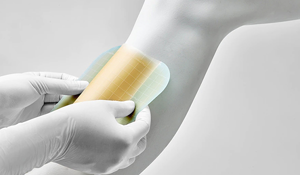
- Hydroactive dressings : multi-layered highly absorbent polymer dressings with a surface adhesive and a waterproof outer layer are similar to hydrocolloids, however, instead of forming a gel in contact with exudate, the fluid is trapped within the product itself, to maintain a moist environment.
- Hydrocolloid dressings : combination of polymers held in a fine suspension. When placed on a wound, the polymers combine with the exudate and form a soft, moist gel-like mass. They also encourage autolysis to aid in the removal of slough from a wound
- Hydrofibre dressings : have some of the properties of alginates in that they are a fibre rope or dressing that forms a firm gel in contact with fluid.
- Foam dressings : soft, open-celled hydrophobic/hydrophilic non-adherent dressings that may be single or multiple layered and meet many of the properties of an ideal dressing
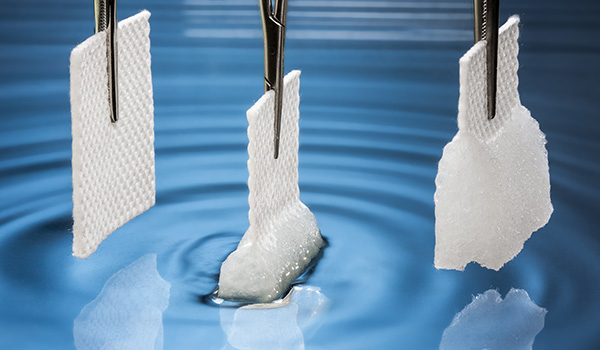
- Alginate absorbent fibre dressings : Alginates are the calcium or sodium/calcium salts of alginic acid, composed of manuronic and guluronic acids obtained from seaweed. When applied to a wound, the sodium salts present in the wound exchanges with the calcium in the alginate to form sodium alginate which is a hydrophilic gel. This gel can absorb exudate into itself while maintaining a moist environment at the interface between the dressing and the tissue.
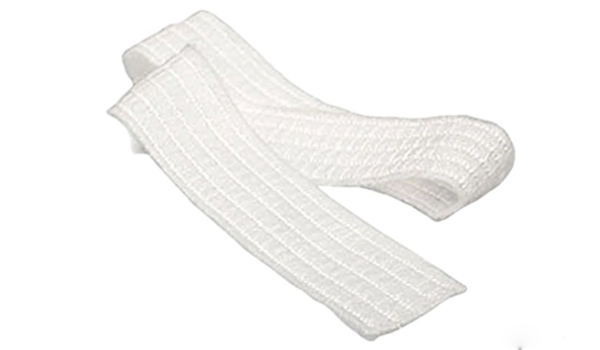
Bandages :
Used from ancient times. In the past 15 years there has been an explosion in the types of bandages available. The bandage may be needed for several reasons:
Retention : keeping a dressing in place
Musculoskeletal support : supporting an injured joint
Compression : assisting venous return from the lower leg.